


توضیحات
معرفی :
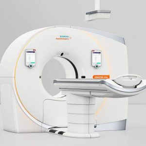
دستگاه سی تی اسکن NeuViz 128 Slice ساخت شرکت Neusoft می باشد که جهت تصویربرداری داخلی از بدن به کار می رود و با عبور پرتو ایکس از بدن بیمار، عکس برداری و دریافت سیگنال ها اقدام به ثبت تصاویر می کند. با استفاده از تصاویر سی تی اسکن می توان بافت های درون بدن را مشاهده کرد و شکل آن ها را مورد بررسی قرار داد. دستگاه سی تی اسکن NeuViz 128 Slice دارای تکنولوژی پیشرفته می باشد که به همین دلیل توانسته استانداردهای بین المللی اروپا CE و آمریکا FDA دریافت نماید و به سراسر جهان صادرات نماید.
دستگاه سی تی اسکن NeuViz 128 Slice با کم کردن هزینه های عملیاتی و استفاده هوشمندانه از تکنولوژی پیشرفته بالینی توانسته است وضوح و دقت قابل توجهی را به تصویربرداری سی تی اسکن هدیه دهد.
دستگاه سی تی اسکن Neuviz 128 Slice ساخت شرکت Neusoft یک دستگاه تصویربرداری پزشکی پیشرفته می باشد که در سال 2017 روانه بازار شد. این دستگاه با بهره گیری از تیوب اشعه ایکس 8MHU و ژنراتور با قدرت 80KW و همچنین سیستم Odose و ClearView که بیان کننده تصویربرداری با کیفیت و دوز پایین اشعه می باشد توانسته قسمت اعظم بازار جهان را از آن خود نماید.
دستگاه سی تی اسکن NeuViz 128 Slice دارای تعداد 64 ردیف دتکتور می باشد و محدوده پوشش دتکتور 40 میلی متر می باشد.
NeuViz 128 Product Highlights
Quad-sampling technology
iHD (isotropic High Definition) imaging enables High Spatial Resolution: 24lp/cm
Micro-STAR detector
1024x1024 large matrix imaging
Comprehensive Low dose design
Clear View, an advanced iterative reconstruction algorithm that adds diagnostic
certainty to low dose imaging
High Resolution Imaging-Chain
Eective integration of high resolution hardware and software results in superior image and diagnostic quality.
Quad-Sampling
By dynamically moving the focal spot axially and longitudinally, sampling density is increased 400%. This means improved resolution, reduced artifact and extended scanning ranges.
Micro-STAR detector
iHD (isotropic High Definition) enables the half-slice acquisition, which delivers
24lp/cm isotropic resolution.
High resolution scanning (1024 x 1024 matrix, small focal spot) provides the spatial resolution necessary to perform lung nodule and inner ear studies.
240 degree exposure
Dose to the patient and attending physician reduced.
Pediatric Protocols
Not “scaled down” adult protocols.
Designed specifically for pediatric anatomy
New detector design
Modular design delivers 99.99% x-ray conversion efficiency,
lower dose necessary to deliver exquisite image quality.
Organ-Safe
Reduces dose to radiosensitive organs such as eyes, thyroid and breasts.
Clear View
Iterative Processing in projection and image spaces that delivers unbelievable dose reduction.
Dose Check
Full implementation of “Dose Check” .
Patient cannot be over radiated.
3D dose modulation
Tube current modulated based on the anatomy in the scan field. Anatomically optimized dose.
ECG dose modulation
Reduces tube current during non-imaging phases of the Cardiac Cycle to minimize patient dose.
| CT Scan 128 Slice | |
| Gantry | |
| 720 mm | Aperture |
| ± 30° | Tilt Range |
| Tube | |
| 8.0 MHU | Heat Capability |
| 931 KHU/min | Heat Dissipation |
| 0.5 x 1.3 mm/1.0 x 1.3 mm | Focal Spot Size |
| Oil to Air | Methode of Cooling |
| Generator | |
| 80 KW | Powering Rate |
| 80, 100, 120, 140 KV | KV Range |
| 10 – 667 mA | MA Range |
| 100 s | Continuous Scan Time |
| Patient Couch | |
| 175 cm | Scan Range / Movement Range |
| 205 Kg | Max Patient Load |
| Console | |
| 0.13 – 2 | Pitch Range |
| 40 images / s | Reconstruction Speed |
| 1024 x 1024 | Image Area Matrix Dimensions |
| Windows 7 | Operating System |
| Detection System | |
| GOS Solid | Detector Type |
| 608 | Number of Detectors |
| 64 | Detector Rows |
| 64 x 0.625 mm | Detector Row Slice Thickness |
| 40 mm | Detector Coverage |
| Helical and Axial Scanning | |
| 0.374 s | Rotation Time For Axial Scan |
| 128 x 0.625, 64 x 0.625, 32 x 0.625, 16 x 0.625, 16 x 0.3125, 8 x 0.625 | Helical Acquisition Widths (Number of Channels x Width mm) |
| Dose Control Technique | |
| O-Dose Auto KV | Low Dose Technique |
| ClearView | Iterative Recon Technique |
| Manufacture’s Performance Data | |
| 0% MTF 24 IP/cm | In Plane Spatial Resolution (Ip/cm) For Sharpest Clinical Algorithm. Acquisition Parameters in Brackets |
| 4 mm @ 0.3%
no more than 39.6 mGy |
Contrast Resolution Smalest Rod Size (mm) Descrenable at Given Parameters in 20 cm CATPHAN |
| Image Storage | |
| >= 1 Tb | Total Standard Hard Disk Capacity (Gb) |
| CD / DVD – RW | Archive Options |
| 3D Reconstruction | |
| Yes | MIPs and MinIPs (maximum and minimum intensity projections) |
| Yes | SSD (3D Shaded Surface Display) |
| Yes | 3D Volume Rendering Software |
| Yes | MPR (Multi Planer Reconstruction) |
| Yes | VR (Volume Rendering) |
| Advanced Applications (MC – main console, WS – workstation) | |
| MC – Standard
WS – Not Available (Bolus Tracking) |
Contrast Media Bolus Tracking |
| MC – Standard
WS – Standard (Vessel Analysis) |
Vessel Analysis |
| MC – Standard
WS – Standard (Cardiac Calcium Scoring) |
Cardiac Calcium Scoring |
| MC – Not Available
WS – Standard (Coronary Analysis) |
Coronary Analysis |
| MC – Not Available
WS – Standard (Cardiac Function Analysis) |
Cardiac Function Analysis |
| MC – Standard
WS – Standard (Nerve System DSA) |
Nerve System DSA |
| MC – Standard
WS – Standard (Brain Perfusion) |
Brain Perfusion |
| MC – Standard
WS – Standard (Body Perfusion) |
Body Perfusion |
| MC – Standard
WS – Standard (Virtual Colonoscopy) |
Colon Analysis |
| MC – Standard
WS – Standard (Lung Density Evaluation) |
Lung Density |
| MC – Standard
WS – Standard (Three Dimensional Lung Nodule Analysis) |
Lung Nodule |
| MC – Standard
WS – Standard (Dental Analysis) |
Dental Analysis |
| MC – Not Available
WS – Standard (Tumor Evaluation) |
Tumor Evaluation |
| MC – Standard
WS – Standard (Fat Analysis) |
Fat Evaluation |


دیدگاهها
هیچ دیدگاهی برای این محصول نوشته نشده است.